
News
Revolutionary imaging technology developed by IBP investigators captured the damage caused by Covid-19 to the lungs’ smallest blood vessels
4 November 2021
Human Organ Atlas
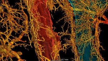
Bronchio-pulmonary shunting in a SARS-CoV-2 infected lung
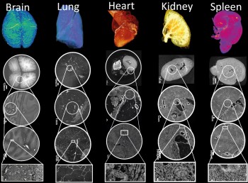
A revolutionary imaging technology called Hierarchical Phase-Contrast Tomography (HiP-CT) developed by our IBP investigator Prof Peter Lee and his team at University College London (UCL) and European Synchrotron Radiation Facility (ESRF). HiP-CT enables 3D mapping the organs across a range of scales, a whole organ level down to down to cellular level. Using HiP-CT, Peter and his team produced a Human Organ Atlas which provides new insights into our biological makeup (please see the paper published in Nature Methods). In addition, with collaboration with clinicians, the team used HiP-CT to gain new insights into how Covid-19 can change our vascular systems (please see the paper published in ATS Journal).
Other resources
Updated by: Ruikang Xue